Clinical Scenario
Native Hip Dislocation
Scenario
A 47-year-old female has had a head on collision with another vehicle at 45mph she was sat in the passenger seat and is now complaining of left hip pain.

Interview Questions
Please interpret the radiograph and tell me what you are concerned about in this patient?
An AP pelvic radiograph is presented in a 47-year-old female. There is evidence of left native hip dislocation. There limb appears to be significantly shortened and in external rotation. There is no obvious associated fracture of the femoral head or acetabulum. I would want further orthogonal views with a lateral radiograph of the hip to assess the direction of dislocation.
Key Concerns
-
High energy injury - ATLS Principles
-
Dislocation requiring urgent reduction
How would you manage this patient?
ATLS
“This patient has a likely high energy injury mechanism; I would therefore ensure that the patient was managed via ATLS principles. With a trauma call, introduction of team members and assignment of roles followed by a primary and secondary survey to identify and treat any life / limb threatening injuries.”
History
-
A Allergies
-
M Medication
-
P Past medical history
-
L Last ate
-
E Events
-
?Previous dislocations
-
Paraesthesia / numbness
-
Other injuries / Pain anywhere else
-
Examination
-
Position of limb
-
Shortened externally rotated - anterior dislocation
-
Adducted, flexed, internally rotated - posterior dislocation
-
-
NV status
-
Examine for sciatic nerve function
-
Investigation
-
XR:
-
AP + Lateral Hip
-
AP Pelvis
-
Management
-
Surgical emergency
-
Initial attempt at Closed reduction in ED with sedation
-
Closed / Open reduction in theatre if unable to perform in ED
-
Escalate to senior
“This patient has a native hip dislocation + paraesthesia. This will need emergency closed reduction in ED under sedation. If unable to reduce, then I would escalate to consultant and book patient into theatre for emergency closed / open reduction.”
You are unable to reduce the hip and your consultant asked you to take patient to theatre overnight for reduction. How would you reduce the hip?
Preparation
-
Explain procedure + gain consent
-
Resus with monitoring
-
Adequate sedation + muscle relaxation by appropriately trained individual
-
Bigelow Manoeuvre
-
Axial traction
-
Internal rotation + adduction
-
-
Allis Manoeuvre
-
Knee flexed to 90 degrees
-
Hip flexed to 90 degrees (relaxes hamstrings)
-
Longitudinal traction in direction of long axis of femur
-
Adduction + internally rotation if required
-
KEY TO REDUCTION = counter-traction from assistant who places down firmly on both ASIS
Note: MUSCLE relaxation may be key to reduction in these patients. Need to ensure discussed with anaesthetist at brief.
Following reduction how would you examine the patient and what imaging would you request?
Post-reduction
-
Check N+V status
-
Check XR
-
AP + Lateral
-
-
CT Pelvis
Why would you request a CT pelvis following native hip reduction?
High energy mechanism, order CT to exclude other injuries:
-
Femoral head # (Pipkin fracture)
-
Acetabular #
-
NOF #
-
Loose bodies in hip joint
What are the complications of a native hip dislocation?
Complications
-
A significant amount of force is required to dislocate a native hip - therefore these are high energy injuries
-
Associated injuries of the acetabulum and femoral head are common and should be assessed for following reduction with a CT pelvis + hip.
-
Associated injuries / complications include:
-
Acetabular fracture
-
Femoral head / NOF#
-
Neurological Injury
-
Recurrent dislocation
-
AVN
-
Due to traction + disruption of retinacular vessels during process of dislocation / reductio
-
-
On examination of the patient how would you differentiate an anterior from a posterior hip dislocation?
Anterior Hip Dislocation
-
Accounts for 10% of dislocations
-
Limb will appear shortened and externally rotated
-
Femoral nerve at risk
Posterior Hip Dislocation
-
Accounts for 90% of dislocations
-
Limb will appear shortened, adducted and internally rotated
-
Sciatic nerve at risk
What neurovascular structures are at risk with an anterior versus posterior hip dislocation?
Anterior = Femoral nerve
Posterior = Sciatic nerve
What injuries must be evaluated for with dashboard type injuries?
-
Posterior hip dislocation
-
Patella #
-
Femoral shaft #
-
Femoral head (pipkin)
-
Posterior wall acetabular fracture
-
Sciatic nerve injury
Following successful reduction of a native hip dislocation - would you follow this patient up in clinic? Why?
Patients with a native hip dislocation should be followed up in clinic for at least 2 years as they have high risk of Avascular Necrosis
-
Due to traction + disruption of retinacular vessels during dislocation
What would be signs that AVN was developing on the follow up radiographs?
Evidence of:
-
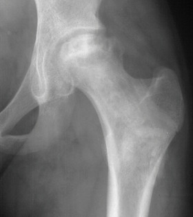
Femoral head sclerosis
-
Fragmentation of femoral head
-
Crescent sign
-
Femoral head collapse
These findings are suggestive of avascular necrosis of the femoral head and may warrant further management such as core decompression or total hip replacement
Note: See image below for AVN femoral head example
Clinical Scenario
Post-operative Foot
Scenario
A 72-year-old male undergoes a THR by a posterior approach. Post-operatively she complains of decreased sensation to the sole of the foot and the physiotherapist notices a foot drop.

Interview Questions
What are you concerned about in this patient and how would you assess this patient?
Key Concerns
-
Sciatic Nerve Injury
-
Need to exclude THR Dislocation - leading to foot drop
History
-
Review operation note
-
Excessive traction intra-operatively
-
Sciatic nerve protected throughout
-
Cement used
-
-
Trauma? Dislocation event?
-
Medications: Anti-coagulation increasing risk of haematoma
-
PMHx
-
Paralysis / paraesthesia
-
Present before operation?
Examination
-
Leg length discrepancies
-
Sciatic nerve stretching with >3cm leg length difference
-
-
?Dislocated hip
-
Shortened, internally rotated = posterior dislocation
-
-
Wound
-
Haematoma?
-
Massive bruising may suggest huge haematoma
-
-
Neurology
-
Check tibial + peroneal divisions of sciatic nerve
-
Investigations
-
Order post-op XR - Exclude hip dislocation
-
CT Pelvis - exclude compressing haematoma
What are the potential causes of sciatic nerve injury following THA?
3 potential Causes
-
Iatrogenic Sciatic Nerve Injury
-
Posterior acetabular retractors
-
Posterior acetabular drill holes
-
Cement extrusion from acetabulum - thermal injury
-
Excessive Femoral Lengthening (>3cm)
-
-
Post-operative haematoma
-
Check for haematoma on exmaination
-
Check if patients on anticoagulants
-
CT to look for compressing haematoma (may need evacuation)
-
-
Post-operative Dislocation
-
Classically posterior dislocation
-
Check for limb length discrepancy and assess position of limb
-
Get an up to date XR to check for dislocation
-
Will require reduction (closed / open) to decompress sciatic nerve
-
Note: Post-operative haematoma can cause compression onto sciatic nerve. Excessive leg lengthening can cause stretch injury of sciatic nerve. Iatrogenic lacerations to the sciatic nerve are rare.
Which division of the sciatic nerve is more commonly injured?
Peroneal division of sciatic nerve is more commonly affected that the tibial division due to its more superficial position
The patient has evidence of an acute post-operative foot drop. How would you manage this patient?
-
Decrease tension on sciatic nerve by flexing knee to 20-30 degrees under pillow
-
Arrange Hip XR to exclude dislocation
-
Inform consultant
-
Keep NBM
-
May need to go to theatre mane for haematoma evacuation and nerve exploration (As per BOAST: Management of peripheral nerve injury [1])
What is Seddon’s Classification of nerve injury?
Seddon’s Classification [2]
Seddon described three basic types of nerve injury:
-
Neuropraxia (Class I)
-
Nerve bruising
-
Focal demyelination usually caused by local ischaemia
-
No Wallerian degeneration
-
Full recovery
-
-
Axonotmesis (Class II)
-
Axon and myelin sheath disruption
-
Endoneurium intact
-
Wallerian degeneration occurs distal to injury
-
Spontaneous recovery possible
-
-
Neurotmesis (Class III)
-
Complete nerve division including endoneurium
-
Wallerian degeneration distal end
-
No spontaneous recovery (unless undergo surgical intervention)
-
References
[1] BOA. BOAST – Peripheral Nerve Injury. Available at: https://www.boa.ac.uk/resources/boast-5-pdf.html
[2] Seddon HJ. A Classification of Nerve Injuries. BMJ 1942;2:237
Images


