Clinical Scenario
Pathological Lesion
Scenario
A 82-year-old male has presented to clinic with increasing right thigh / hip pain. He has no history of trauma and reports weight loss of 1 stone over the past 3 months.

Interview Questions
Please interpret the radiograph and tell me what you are concerned about in this patient?
An AP radiograph of the right proximal femur is presented in an 82-year-old male. There is a lytic lesion at the junction of the proximal to middle third of the femur with cortical involvement. There is a narrow zone of transition with no periosteal reaction visible. This lesion is suspicious for a pathological lesion and requires further work up.
Key Concerns
-
Atraumatic hip fracture = Needs investigation for pathological fracture
-
Manage as per BOAST: Management of Metastatic Bone Disease [1]
What questions would you ask in this patient’s history? How would you examine them?
History
-
Pathological lesion Questions
-
Preceding bone pain
-
Weight loss, fevers, night sweats
-
Systems review:
-
Thyroid - Hoarseness? Neck lumps?
-
Breast - Lumps?
-
Lung - SOB? Haemoptysis? Cough
-
Prostate - ?difficulty urinating
-
Kidney - ?haematuria
-
-
-
Previous bisphosphonate usage
-
Past medical history / social history (smoker)
Examination
-
NV intact?
-
Thyroid / breast / prostate / kidney / lung examination
-
Inguinal lymphadenopathy
-
Spinal Tenderness
Note: These patient's need full oncological history and examination work up. Should examine for 5 common areas for primary bone tumours.
What investigations would you order?
Investigations
-
Bloods
-
FBC, U&Es, CRP, LFTs, Clotting
-
Bone profile
-
(Raised Alkaline Phosphatase / Hypercalcaemia may raise suspicion of malignancy)
-
-
Thyroid Function Tests (TFTs)
-
Prostate Specific Antigen (PSA) + Myeloma screen
-
-
Imaging
-
Xrays
-
Lateral view Hip
-
AP Pelvis
-
Full length-femur
-
-
Specialist imaging:
-
CT CAP
-
Should be performed within 24 hours (as per BOAST)
-
-
MRI
-
Bone Scan
-
-
If you were suspecting a primary bone tumour, what would you do? What about management if suspecting a secondary bone tumour?
Management depends on whether there is a primary or secondary tumour
Primary Tumour
-
Refer to regional sarcoma unit
-
Discuss at Multi-Disciplinary Team (MDT) meeting
-
Bone biopsy to be performed at regional centre - usually undertaken by operating surgeon due to risk of seeding tumour along the biopsy track
Secondary Tumour
-
Discuss at MDT
-
Is solitary bone metastasis then should be referred to teritary referral unit (as per BOAST)
-
Consider prophylactic fixation (e.g. IM Nailing) if impending fracture
-
Other treatment options include radiotherapy / chemotherapy
What patients should be referred to a tertiary referral unit?
As per BOAST guidance on Management of Metastatic Bone disease. The following should have referral onwards to a tertiary referral unit:
-
Primary bone tumours - within 72 hours
-
Solitary bone metastases
Metastatic disease without an obvious primary site should be discussed with acute oncology services
What are the most common primary sites for metastatic bone lesions? Which of these are classically lytic? Sclerotic? Mixed?
Metastatic Disease
Most Common Primary Sites
-
Thyroid
-
Breast
-
Lung
-
Kidney
-
Prostate
Appearance
-
Sclerotic (blastic) = Prostate
-
Mixed = Breast
-
Lytic = Thyroid / Renal / Lung
If a patient had a renal cell carcinoma metastasis, what might you consider prior to surgical fixation?
Renal Cell Carcinoma metastases are extremely vascular and pre-operative embolization should be considered to reduce risk of bleeding during fixation
What criteria can be used to predict need to perform prophylactic fixation in metastatic bone lesions?
Mirels’ Criteria [2]
-
Predicts risk of pathological fracture in patients with long bone metastases
-
Can be used to help decision whether to perform prophylactic fixation
-
Consists of four separate criteria with a score of 1-3
-
(3 indicating higher risk of fracture)
-
Addition of 4 criteria numbers gives overall risk
-
-
A score of greater than or equal to 8 = prophylactic fixation
What are the aims of surgery in metastatic bone cancer treatment?
-
Survive surgery
-
Allow Immediate full weight bearing
-
Implant should outlive patient
-
Protect entire length of bone
-
Pain relief
"BOAST guidelines recommend that surgery should be consultant-led, aim for long-term durability, and allow immediate weight-bearing.
If performing a prophylactic IM nail fixation on this patient - what should be done prior to nail insertion?
Venting of the IM canal when performing IM nailing – can reduce the risk of fat embolization
Describing a bone tumour on Radiographs
-
Location - Which bone? / Where abouts in the bone? (Diaphyseal Vs metaphyseal)
-
Size
-
Transition Zone - Narrow Vs wide
-
Periosteal Reaction
-
Cortical Involvement
-
Matrix Appearance - Osteoid Vs Chondroid
What is the definition of subtrochanteric fractures?
Definition: Typically defined as an area from the lesser trochanter to 5cm distal to the lesser trochanter
When presented with a subtrochanteric fracture need to exclude:
-
Pathological Fracture OR
-
Bisphosphonate Fracture
What are the features of bisphosphonate related fracture on radiographs?
Bisphosphonate Related Fractures
-
Around 80% of subtrochanteric fractures are associated with long-term bisphosphonate usage
-
These fractures are associated with increased healing times
-
Fractures fail in tension - starting at lateral aspect of femur - then spreads to medial aspect causing cortical beaking
What are the features of bisphosphonate related fracture on radiographs?
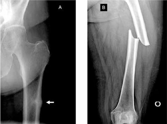
Classical Features on Radiograph (see images below)
-
Increased cortical thickening
-
Transverse / short oblique fracture orientation
-
Medial Spike = cortical beaking
-
Lack of comminution
References
[1] BOA. BOAST- Management of Metastatic Bone Disease. Available online at: https://www.boa.ac.uk/resource/boast-management-of-metastatic-bone-disease.html
[2] Mirels H. Metastatic disease in long bones: A proposed scoring system for diagnosing impending pathologic fractures. Clin Orthop Relat Res. 2003;415:S4-13
Images


